Frozen Shoulder: What the World’s Largest Clinical Trial Reveals About Surgery vs. Therapy12/5/2026
For those living with "frozen shoulder"—medically known as adhesive capsulitis—the world shrinks to the radius of a locked joint. It begins with an insidious onset of deep-seated pain that gradually hardens into a debilitating "gridlock." Simple gestures become Herculean tasks: reaching for a seatbelt, pulling on a coat, or finding a sleeping position that doesn't trigger a jolt of agony. In this state of inflammation and scarring, the shoulder joint contracts, leaving patients desperate for a solution that will restore their range of motion.
To settle the debate over which intervention truly works, the UK FROST study was launched. As a multicentre, pragmatic, three-arm, superiority randomised clinical trial—the largest of its kind—it put the three most common secondary care treatments to the ultimate test. The study aimed to determine whether expensive, invasive surgery is actually superior to a structured physiotherapy pathway. The Superiority Myth: All Roads Lead to Recovery The most striking revelation from the UK FROST study was that at the 12-month mark, no single treatment proved "clinically superior." To measure success, researchers used the Oxford Shoulder Score (OSS), a 48-point scale where higher scores indicate better function and less pain. The trial was designed with a "target difference" of 4 to 5 points—the minimum improvement a patient would actually notice in their daily life. While the results showed that patients who underwent Arthroscopic Capsular Release (ACR) had statistically better scores than those in the physiotherapy group (a difference of 3.06 points, p=0.011), the gap failed to reach that critical 5-point clinical threshold. In the language of evidence-based medicine, this is a vital distinction: the surgical advantage was "statistically significant" (meaning it likely wasn't due to chance), but it wasn't "clinically significant" (meaning the patient wouldn't feel a meaningful difference between the two). As the study authors concluded: "none of the three interventions were clinically superior." Whether the patient chose the scalpel or the exercise mat, the 12-month outcome was remarkably similar. The Cost of Invasive Action: Safety and the Surgical Scalpel While clinical outcomes were nearly identical at one year, the journey to get there varied significantly in terms of risk. The trial evaluated three distinct pathways:
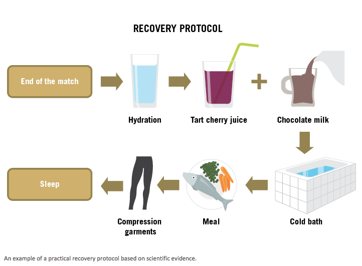
The safety data was telling. ACR carried the highest risk profile, with eight serious adverse events reported, including one patient suffering a stroke and others experiencing deep vein thrombosis or surgical site infections. In contrast, MUA saw only two serious events, and the early structured physiotherapy group saw zero. This suggests that while ACR is a powerful tool, its higher complication rate makes it a "selective" option rather than a default first-line treatment. The Waiting Game: Why Access is Therapy For a patient whose life is on hold, the most important metric isn't just how they recover, but how fast they can start. The UK FROST study revealed a massive disparity in access. The median wait time for physiotherapy was just 14 days. For MUA, it was 57 days, and for ACR, it stretched to 72 days. This delay has real-world consequences. At the 3-month follow-up, the ACR group actually reported worse outcomes than the other two groups. This "surgical lag" occurred because many ACR patients were either still on the waiting list or were in the early, painful stages of post-operative recovery while the physiotherapy group had already completed their treatment. When you are unable to sleep or work, a two-month head start on recovery is a significant clinical advantage. Efficiency in the Theatre: The Economic Winner From a healthcare system perspective, the UK FROST study provides a clear economic winner: Manipulation Under Anaesthesia (MUA). The researchers used Quality-Adjusted Life-Years (QALYs)—a metric where one unit represents one year of perfect health—to determine value for money. At the standard NHS threshold of £20,000 per QALY, MUA had an 86% probability of being the most cost-effective treatment. ACR, by comparison, was substantially more expensive—costing roughly £1,733 more per patient than physiotherapy—without providing a commensurate leap in health quality. In a system where hospital beds and operating theatre time are precious resources, MUA offers the most efficient balance of cost and clinical improvement. The Reality of Persistence There is a final, sobering takeaway for both clinicians and patients. While the treatments in this trial were highly successful—most participants reached nearly full function with a median score of 43 out of 48—frozen shoulder remains a stubborn adversary. Historical data on the general population suggests that around 40% of patients may still report some persistent symptoms even four years after the initial onset. While the UK FROST participants generally fared better, the "slow or incomplete" nature of recovery in the broader population serves as a reminder that this condition is a marathon, not a sprint. There is, however, a notable trade-off regarding further intervention. While the physiotherapy pathway is safer and faster to access, 15% of those patients eventually required further treatment (such as surgery) compared to only 4% of those who started with ACR. A Blueprint for Future Shoulder Care The UK FROST study has effectively redrawn the map for adhesive capsulitis treatment. It proves that more invasive does not necessarily mean better. For the patient in the consultation room, the "Early Structured Physiotherapy" pathway—the combination of a steroid injection followed by expert-led exercise—should be a primary consideration. It is fast, safe, and at 12 months, delivers results that are nearly indistinguishable from surgery. MUA stands as the most cost-effective hospital intervention, while ACR is best reserved for complex cases or when less invasive methods have failed. Ultimately, the study empowers the patient. If the long-term outcomes are virtually the same, would you choose the surgical theater and the risks that come with it, or would you choose the injection and the exercise mat? The evidence suggests that for most, the less invasive path is just as effective. REF: The Cold, Hard Truth: What Science Actually Says About Your Post-Race Recovery 1. Introduction: The Perpetual Plateau For the dedicated endurance athlete—the marathoner, the triathlete, the cyclist covering triple-digit mileage—recovery is often viewed as a mathematical equation: investment equals adaptation. Yet, despite the billions spent on pneumatic sleeves, massage guns, and boutique supplements, the "perpetual plateau" remains a common frustration. You finish your session, deploy your tech, and still wake up with the dreaded "heavy legs." This isn't just an inconvenience; it’s a performance risk. As a consultant, I look at the stakes: an imbalance between training stress and recovery doesn't just stall your PRs—it leads to metabolic disturbances, systemic inflammation, and the very real danger of non-functional overreaching (NFO). To cut through the marketing noise, we must look at the 2024 "Umbrella Review" by Li et al., the first global synthesis of recovery data specifically filtered for endurance athletes. The core question remains: if we’re doing more for recovery than ever before, why does the data suggest we are largely missing the mark? 2. Takeaway 1: The "Magic Bullet" Does Not Exist The most jarring reality of the Li et al. review is found in its "donut charts" of data. Across almost every recovery modality, the dominant finding was "No Effect." The researchers analyzed ten distinct parameters—including biochemical markers like Creatine Kinase (CK), performance metrics like Time to Exhaustion (TTE), and biomechanical variables—and found that no single strategy consistently moved the needle across the board. What works for one marker (reducing subjective muscle soreness) often does absolutely nothing for another (restoring VO2max or jump power). This is why a "blind" recovery protocol is a failing protocol. Fixing a biochemical marker like CK doesn't mean your neuromuscular system has "reset" for tomorrow’s intervals. As the study explicitly concludes: "There is no particular recovery strategy that can be advised to enhance recovery between training sessions or competitions in endurance athletes." 3. Takeaway 2: Why Endurance Athletes Are Not Team Players Generic recovery advice is often "contaminated" by data from team sports like soccer or basketball, where the physiological demands are fundamentally different. To understand the "different league" endurance athletes inhabit, look at the Metabolic Equivalent (MET) hours. While bodybuilding sits at a 6.0 and basketball at 8.0, marathon running, triathlons, and speed skating demand a staggering 13.3 MET hours. Even rowing (12.0) significantly outpaces the 10.0 MET hours seen in professional soccer. When a elite marathoner is covering 150–260 km per week, they are inducing a level of metabolic disturbance that renders "standard" recovery advice useless. We cannot apply the recovery needs of a power-based athlete to a person whose primary stress is submaximal intensity for prolonged durations. 4. Takeaway 3: The Massage Myth—Feel Good vs. Function Massage remains the most popular recovery tool in the endurance community, but from a performance consulting perspective, it is largely a "psychological placebo." The review found its effects on objective physiological markers—lactate clearance, VO2max, and heart rate—to be "marginal or nonexistent." Crucially, the data shows that massage is actually less effective for trained endurance athletes than for untrained individuals or those engaging in high-intensity "mixed" exercise. For the 8–24 hour training recovery window, studies found zero benefit from manual or vibration massage on actual performance output. It makes you feel better by addressing perceived soreness (DOMS), but it does not prepare your muscles to function at a higher capacity the next day. 5. Takeaway 4: The Promising Duo—Compression and Cold If there is a light in the "messy" data, it shines on Compression Garments (CG) and Cryotherapy. These were the only two strategies identified as "promising" for the critical 8–24 hour Training Recovery window—the phase where actual physiological adaptation occurs.
While CWI is often criticized for blunting hypertrophy in resistance training, the cold appears to be a genuine ally for the endurance athlete focused on maintaining high-volume performance between sessions. 6. Takeaway 5: Active Recovery Beats the Couch The Li et al. review enforces a vital conceptual shift pioneered by researchers like Kellmann: Rest is inactivity, but Recovery is an additional stimulus. The data compared active recovery (voluntary submaximal movement) against "seated rest." For swimmers and climbers, 6–10 minutes of submaximal activity resulted in significantly better lactate clearance. By viewing recovery as a light, intentional movement stimulus rather than "couch time," you facilitate the clearance of metabolic waste and maintain the body's readiness for the next training load. 7. Conclusion: A Forward-Looking Framework The scientific reality of endurance recovery is a landscape of individualisation. We currently face a "proactive gap"—we lack high-quality, endurance-specific data on the biggest pillars of performance, namely sleep and alcohol consumption. Until that data matures, the most effective framework is to focus on the 8–24 hour Training Recovery window using proven tools: cold exposure, compression, and active movement. Everything else is likely just "feeling" good. Final Thought: In a world of expensive gadgets, are you prioritizing the psychological "feel" of a massage over the physiological "function" of 10 minutes of active movement and a pair of compression socks?
The Soccer Header Dilemma: Why the Science on Youth Safety Is Surprisingly Unsettled In 2021, England Football implemented a sweeping set of guidelines that effectively removed heading from training for children under 11. This move was rooted in the "precautionary principle"—the idea that it is better to restrict a behavior now than to wait for definitive proof of harm later. However, this policy rests on a surprisingly thin foundation; we are regulating a "safe dose" of head impacts without actually knowing what that dose is, or if the impacts are truly damaging. The central tension in youth sports today is the gap between these rapid policy changes and the unsettled nature of the underlying science. While the public remains focused on "subconcussive" hits—impacts that don't cause immediate symptoms—the data suggests our anxieties may be directed at the wrong target. As we move toward more restrictive rules, we risk altering the fabric of the sport based on fears rather than firm evidence. The Surprising Statistical Safety of the Head vs. the Leg Statistically speaking, a child’s head is one of the safest parts of their body on a soccer pitch. Research indicates that acute head and neck injuries in youth football occur at a rate of just 0.25 per 1,000 hours played. This pales in comparison to lower leg injuries, which occur at a rate of 4.08 for males and 6.54 for females per 1,000 hours. This disparity reveals a significant "unintended consequence" of current policy. By banning heading to stop subconcussive hits, we also reduce "aerial competitions"—the moments when two players jump for the same ball. These competitions are the primary source of dangerous head-to-head or head-to-extremity collisions. Because the science hasn't yet separated the risks of purposeful heading from these accidental collisions, we may be over-regulating the header while ignoring the broader context of how concussions actually happen. The "Developing Brain" Sensitivity Hypothesis The urgency for youth guidelines stems from the belief that a developing brain is more susceptible to long-term damage than an adult brain following mild injury. While adult studies show mixed results, some have identified acute increases in corticomotor inhibition (a temporary suppression of the brain’s signaling to muscles) and decreased memory performance after heading. To manage this, adult professional players in the UK are now limited to just 10 "high force" headers—such as those from crosses or long passes—per training week. Governing bodies are applying similar logic to children, even though the evidence for long-term harm from purposeful heading remains uncertain and under-researched. The goal is to eventually move past guesswork and establish a scientifically validated "maximal safe dose." By investigating how these impacts affect neurodevelopment, researchers hope to identify specific risk factors that make some players more vulnerable than others. The Playground Policy Gap While professional academies can meticulously log every impact, the "majority" of youth soccer happens in an informal world beyond the reach of any governing body. In schools, parks, and backyards, there are no coaches to enforce England Football’s U11 restrictions or monitor heading frequency. This creates a massive "playground gap" where the most well-intentioned training rules fail to account for the total volume of head impacts a child receives. Policing a mass participation sport is notoriously difficult, particularly when the play is unorganized. If the goal of policy is to limit cumulative exposure, the current focus on "official" training sessions may only be scratching the surface. Without a way to monitor the informal game, these safety rules might provide a false sense of security while the actual "dose" of impacts remains unrecorded. The Data Deficit and the Danger of "Assuming the Worst" The current evidence base for heading restrictions is remarkably small, with many studies relying on fewer than 20 participants. Much of the public concern is actually extrapolated from American Football data, where the frequency and force of impacts are vastly different. This "preponderance of data" from a different sport makes it difficult for soccer governing bodies to recommend sweeping, evidence-based changes that are specific to the unique mechanics of the world’s most popular sport. There is a real risk that if public fear outpaces scientific reality, the sport will suffer unnecessary damage. Clinicians warn that we must balance "unproven risks" against the clear, documented advantages of team sports. "The goal of researching the potential neurological harms... is not to dissuade young people from playing the sport. The goal is to better understand the difference between purposeful heading and concussion... and to elucidate factors that may exacerbate one’s risk of developing neurological impairments (e.g., height, strength, position played)." A Data-Driven Path Forward To preserve the sport, we need high-quality research that moves beyond the precautionary principle and toward a true risk-benefit assessment. Safety can be improved through practical, technical alterations rather than just bans. This includes using age-appropriate equipment, reducing the weight and pressure of balls, and limiting "long balls" over 35 meters in training to reduce high-force impacts. Focusing on the technique of aerial competitions, rather than just the act of heading, could address the most dangerous collisions while keeping the game intact. We must be mindful of the plethora of societal benefits—from cardiovascular health to social interaction—that soccer provides. The challenge for the next decade is to ensure that our safety rules are built on the firm ground of quality research, ensuring the game remains both safe and enjoyable for the next generation.
The Strength Paradox: Why Exercise Heals Your Pain (Even When You’re Not Getting Stronger)
Imagine a dedicated runner who has spent months in the gym treating a nagging knee. They have followed every protocol, their quads are visibly larger, and they are lifting heavier weights than ever before—yet the pain persists with every stride. This frustrating scenario highlights the "Strength Paradox": the assumption that physical weakness is the root of pain, and that muscle growth is the only path to a cure. While exercise remains the gold standard for managing musculoskeletal (MSK) pain, recent clinical data suggests we have been misidentifying the "why." We are discovering that while exercise heals, the "mechanic" behind the recovery is rarely the muscle itself. It is time to bridge the gap between what we believe about our bodies and what the research actually reveals. The "Chasm" Between Belief and Data For decades, the medical community has operated under a biomechanical lens, assuming a direct "causal mediation" between muscle power and pain relief. We tell ourselves that pain diminishes because strength increases. However, when researchers look for the actual link between these two variables, the evidence is surprisingly thin. This mismatch creates a significant hurdle for both clinicians and patients. When we rely on outdated beliefs rather than mechanistic evidence, our treatments become less stable and harder to justify. As the source material highlights, the discrepancy between our clinical assumptions and the actual data is no longer something we can ignore. "The chasm between belief and data warrants attention." Takeaway 1: Your Muscles Aren't the Problem (The Evidence) The most striking evidence against the "strength-as-cure" model comes from studies on tendons and shoulders. In many cases, patients experience profound relief from pain and disability without any corresponding change in their muscle structure or power. This suggests that while strength might improve during a program, it isn't the cause of the relief. The lack of a causal link is documented across several common conditions:
Takeaway 2: The 2% Reality Check In the world of knee Osteoarthritis (OA), the data is even more humbling. An individual participant data meta-analysis found that knee extension strength accounted for only about 2% of the treatment effect of exercise. For a patient told their pain is due to "weak knees," this statistic is a revelation. If strength is only responsible for 2% of the benefit, then 98% of why exercise works remains a "black box" in traditional biomechanics. This suggests our clinical focus is almost entirely misplaced. When we obsess over muscle power, we are ignoring the massive, hidden drivers that actually facilitate healing. Takeaway 3: It’s Not Just Mechanical—It’s "Bio-Psycho-Social" If strength isn't the primary mediator, what is? Evidence points to a "constellation" of biological, psychological, and social factors. These mechanisms are shaped by "contextual factors" like the patient's personal goals, values, and preferences, all of which are optimised by a strong therapeutic relationship between patient and clinician. Pain Self-Efficacy and Beliefs This is the confidence in your ability to move and function despite having pain. When exercise helps you realize that your body is capable and resilient, your disability levels drop. Altering your beliefs about what your body can handle is often more curative than any muscle contraction. Reducing Kinesiophobia and Catastrophising Exercise is a powerful tool for reducing "kinesiophobia" (fear of movement) and "pain catastrophising" (dwelling on the worst-case scenario). In back and shoulder pain, exercise works by teaching the brain that movement is safe. Once the fear and the expectation of disaster are removed, the pain experience often softens. Biochemical Homeostasis Movement triggers internal "housekeeping" at a microscopic level. In conditions like knee OA, exercise combined with diet can reduce inflammatory biomarkers and improve the internal environment of the joint. This biochemical shift provides relief that has nothing to do with the size of the surrounding muscles. Takeaway 4: Why "Weakness" is a Dangerous Narrative Understanding the how of exercise isn't just an academic exercise; it is a matter of scientific rigour, clinical integrity, and responsible research. When we tell patients they are "weak" or "fragile," we risk demoralizing them if they don't see immediate physical gains. This narrative can reinforce harmful, protective behaviours that actually slow down recovery. We must shift the conversation toward "building confidence and adaptability." If clinicians only report the total effect of exercise without understanding the mediators, they miss the opportunity to design more efficient treatments. Professional integrity requires us to stop using "fixing weakness" as a catch-all explanation and instead help patients understand their body's incredible capacity to adapt. Takeaway 5: Why We Should Keep Lifting Anyway Does this mean we should stop lifting weights? Absolutely not. Strength is still a vital biomarker for general health, longevity, and reducing fall risks in older adults. It is also a key indicator of functional success after specific procedures, such as ACL reconstructions. The goal is to change the narrative. We shouldn't lift just to "fix" a specific pain point; we lift to increase our physical robustness and overall health. As we move away from the "weakness" myth, we can offer a more honest and empowering message to those in pain. "Exercise can benefit many people with MSK pain, even when underlying mechanisms vary or remain unclear. Through keeping active and gradually challenging your body, exercise can help you adapt in ways that build confidence, reduce pain and improve function, ultimately helping you get back to doing the things you want and need to do. The key is finding the type of exercise that works for you, your goals, your experience and your interests." Conclusion: A New Framework for Moving Forward Recovery is a complex, multidimensional journey that cannot be reduced to a single muscle measurement. By moving away from the idea that we are "broken" or "weak," we embrace a framework of physical robustness that respects the brain, the immune system, and the person as a whole. The goal of movement is to help your body adapt and thrive in an uncertain environment. If your recovery isn't just about the strength of your muscles, how much more freedom do you have to find a movement you actually enjoy? REF: It is not all about strength: rethinking mechanistic assumptions in exercise-based rehabilitation for musculoskeletal pain relief
The ACL Myth: Why Your First Move After a Tear Might Not Be the Operating Room
It is a sound that haunts every athlete: the sickening "pop" followed by immediate instability and the realization that your Anterior Cruciate Ligament (ACL) has given way. For decades, the script following this injury has been nearly universal—schedule surgery, undergo reconstruction, and begin the long road to recovery. The assumption was that without a surgical fix, a "stable" knee and a return to a normal, active life were impossible. However, a groundbreaking "living" systematic review and meta-analysis recently published in the British Journal of Sports Medicine is shifting this narrative. What makes this a "living" review is its commitment to the cutting edge; the researchers plan to update their findings every year as new data emerges. Currently, their synthesis of randomized controlled trials (RCTs) is challenging the "surgery-first" status quo, suggesting that for many, the immediate trip to the operating room may not lead to better results than a dedicated rehabilitation program. The Functional Dead Heat When patients choose surgery, they are usually looking for one thing: a knee that feels and performs like it did before the injury. Researchers measured this using self-reported scores, such as the International Knee Documentation Committee (IKDC) and the Knee Injury and Osteoarthritis Outcome Score (KOOS). While the current evidence is considered low to very low certainty, the study found no clinically important differences in knee function between those who had early surgery and those who started with primary rehabilitation. This parity held true across short, medium, and long-term follow-ups. It is a striking finding: despite the invasive nature of reconstructive surgery, patients’ perceived quality of movement and stability was nearly identical to those who opted for the gym over the scalpel. "Current evidence suggests that both early surgery and primary rehabilitation result in clinically meaningful improvements in long-term subjective knee function." The Arthritis Paradox One of the most enduring arguments for early ACL surgery is that it "protects" the knee from future wear and tear, specifically knee osteoarthritis (OA). The logic seems sound—stabilize the joint mechanically to prevent the bones from grinding. However, the data tells a different story. The review found that early reconstruction showed no protective effect against osteoarthritis. In fact, primary rehabilitation showed a positive trend for better radiological outcomes—meaning better results on X-rays and MRIs—though the certainty of this evidence remains very low. The researchers identified four potential reasons why surgery might fail to prevent, or could even contribute to, joint degradation:
"Our findings from RCTs challenge a historical paradigm that anatomic instability must be stabilised with surgery to prevent knee osteoarthritis." The "Stepped Care" Revolution Rather than viewing surgery and rehab as a binary choice, the study advocates for a "stepped care approach." In this model, high-quality, supervised rehabilitation is treated as the first-line treatment for most ACL patients without serious concomitant injuries (such as repairable meniscal tears or other high-grade ligament damage). Under this framework, surgery is reserved as a secondary option specifically for the subset of patients who experience persistent "functional instability" despite their efforts in rehab. This approach allows many patients to avoid the inherent risks of the operating table altogether while improving the overall cost-effectiveness of care. It shifts the focus from "surgery for all" to a strategy where the scalpel is only used when the strength gained in the gym hasn't solved the functional problem. The Meniscal Catch: When Waiting Becomes a Risk While functional outcomes were similar, the study did find some nuance regarding the meniscus—the knee’s shock-absorbing cartilage. There was a slight trend, albeit with low certainty, suggesting that early surgery might lead to better meniscal outcomes in the long run. Crucially, the "as-treated" analysis revealed that the worst outcomes were often seen in the "delayed surgery" group—those who attempted rehab but eventually required surgery due to persistent instability. This highlights the necessity of "shared decision-making" between the patient and clinician. Individual factors, such as a patient's unique tibial slope or high functional demands, must be weighed carefully to determine if they are a strong candidate for a "rehab-first" track or if their specific anatomy requires early stabilisation to protect the meniscus. Why "Return to Sport" Isn’t a Guarantee for Either Side Many athletes rush to surgery because they believe it is the only ticket back to the field. Using the Tegner Scale to measure activity levels, the researchers found that for the average person, neither treatment was significantly superior for returning to pre-injury activity. However, the "smart-friend" truth is a bit more sobering: the review notes that while many athletes return to sport, many do not reach their pre-injury level of performance, regardless of whether they chose surgery or rehab. Furthermore, while the trend shows no difference for the general population, we still lack high-quality data specifically focusing on "extreme high-level" professional athletes (Tegner level 10). For the rest of us, the data—though currently of very low certainty—suggests that neuromuscular control is just as vital as a new ligament for getting back to the game. A New Way to Heal The debate between the "scalpel and strength training" is evolving into a more nuanced conversation. We are moving away from a one-size-fits-all surgical mandate toward individualized, patient-centered care. While the certainty of the current evidence remains low to very low, it suggests that for many, a focused rehabilitation program can yield the same functional quality of life and potentially better long-term joint health than immediate surgery. Ultimately, the decision to undergo surgery should be a collaboration, not a foregone conclusion. If the outcomes are the same, would you choose the risk of the operating table or the hard work of the gym? REF: Primary surgery versus primary rehabilitation for treating anterior cruciate ligament injuries: a living systematic review and meta- analysis
Practical recommendations on stretching exercise: A Delphi consensus statement of international research experts
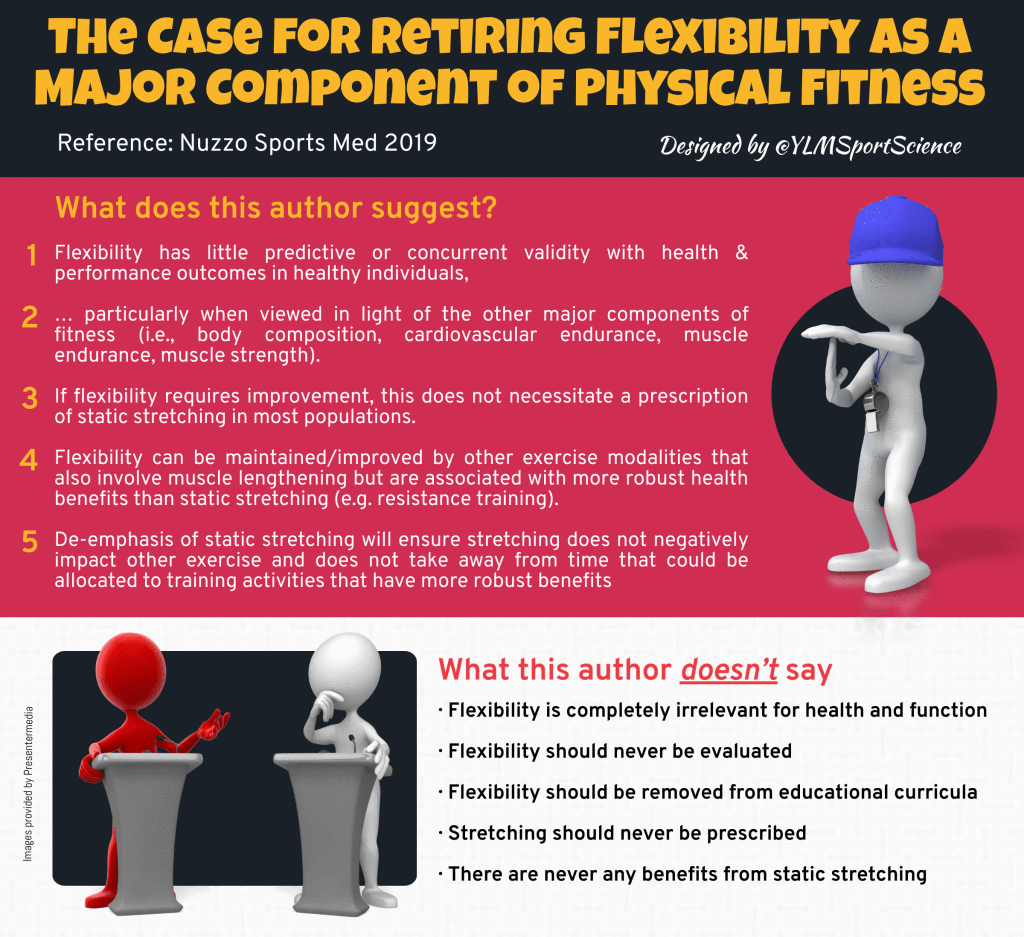
For decades, stretching has been the ultimate fitness ritual. Whether you are a weekend warrior or an elite athlete, reaching for your toes is often viewed as a mandatory prerequisite for performance and a non-negotiable insurance policy against injury. But as our understanding of sports science evolves, we must ask: does this ritual actually deliver on its promises? The 2025 International Delphi Consensus recently set out to reconcile years of conflicting data. A panel of 20 international experts from 12 countries meticulously reviewed the evidence to provide a new roadmap for movement. Their findings challenge "common wisdom," suggesting that while stretching has unique benefits, it may not be the panacea we once believed. The "60-Second Rule" for Performance One of the most significant findings involves the "performance killer" effect of static stretching on strength. The experts reached a 95% consensus that holding a stretch for too long immediately before explosive movement can hinder results. This impairment is primarily observed in isolated muscle groups subjected to maximal or explosive contractions. For example, prolonged static stretching of the calves may decrease your vertical jump, but it is unlikely to impact your bench press. The consensus establishes a clear threshold: avoid static holds longer than 60 seconds per muscle group if your goal is immediate power. Short-duration stretches integrated into a dynamic warm-up remain a safe, viable option. "The panel does not recommend prolonged (>60 s per muscle) static stretching prior to maximal or explosive contractions in isolated muscle groups." Stretching for Hypertrophy: The High-Intensity Requirement A surprising takeaway is that stretching can actually build muscle mass and strength, reaching a 90% consensus among experts. However, this isn't the "passive" relaxation most people imagine. To trigger structural growth, the protocol requires High-Intensity Static Stretching (HI-SS), which is significantly more demanding than traditional flexibility work. This "high-dosage" approach is particularly valuable for "clinical populations" or elderly individuals who are unable or unwilling to perform traditional resistance training. It serves as a minimalist entry point for those currently sedentary. To achieve measurable hypertrophic gains, the panel identified specific requirements:
The Injury Prevention Paradox The belief that stretching is a universal shield against injury was met with scientific skepticism. The panel reached an 85% consensus that stretching is not an "all-encompassing" prevention strategy. While some evidence suggests static stretching might reduce the incidence of muscle-specific injuries, it introduces a documented trade-off. Data from recent systematic reviews indicate that a reduction in muscle injuries may be balanced by an increase in bone and joint injuries. This paradox suggests that athletes must carefully weigh the marginal benefits of stretching against the potential risks and the time invested. For many, stability and strength training remain more efficient interventions for overall safety. The Recovery Myth: DOMS vs. Reality Many of us reach for a post-workout stretch to ward off Delayed Onset Muscle Soreness (DOMS). However, the panel reached a 100% consensus that stretching does not substantively improve post-exercise recovery compared to passive rest. It fails to restore strength or range of motion faster than simply doing nothing. This finding aligns with a shift in physiological understanding: DOMS is now believed to originate in the deep fasciarather than the muscle fibers themselves. Because traditional stretching does not address these underlying fascial or inflammatory processes, it remains a matter of individual psychological preference rather than a physiological necessity. Stretching as a "Vascular Therapy" The most impactful "future-facing" insight from the consensus concerns the circulatory system. The experts identified stretching as a viable "vascular therapy" reaching a 90% to 95% consensus for acute and chronic benefits, respectively. Chronic stretching has been shown to improve heart rate variability and reduce arterial stiffness. For immediate (acute) vascular improvements, the panel recommends a minimum of 7 minutes per muscle. For long-term (chronic) cardiovascular health, the protocol mirrors the hypertrophy dosage of 15 minutes per muscle. This makes stretching a powerful alternative for those currently unable to engage in active cardiovascular exercise. "The panel agrees static stretching could be an alternative for those unable to engage in active (therapeutical) exercise... to reduce arterial stiffness, increase heart rate variability, and improve endothelial function." Posture Cannot Be Stretched into Alignment The consensus took a direct stand against the idea that stretching alone can "fix" poor posture. In the past, practitioners often recommended stretching the "tight" chest to pull the shoulders back. The panel reached a 100% consensus that isolated stretching is ineffective for changing spinal alignment or conditions like Upper Crossed Syndrome. The experts suggest viewing stretching as an "incomplete tool" for postural correction. While isolated stretching fails, a combined approach that partners stretching with the strengthening of weak muscles has shown efficacy. If you seek to change your alignment, strengthening is the essential partner that stretching cannot replace. The Verdict: A New Way to Move The 2025 Delphi Consensus reveals that many flexibility gains are actually due to increased stretch tolerance—the brain's ability to handle discomfort—rather than actual structural tissue elongation. This distinction shifts stretching from a mandatory "ritual" to a strategic choice. It is a viable tool for specific goals like range of motion or vascular health, but it is rarely a necessity. As we move forward, the focus must shift from tradition to intent. If stretching is a choice rather than a requirement, how will you choose to spend your limited gym time? When the ritual is stripped away, the question remains: are you moving with purpose, or simply following a habit the evidence no longer supports?
We are currently facing a mental health crisis of staggering proportions. Globally, depression and anxiety affect between 7% and 25% of the population, leaving millions searching for relief. Perhaps most alarming is that our youth are experiencing these conditions at twice the rate of adults, a trend that threatens the well-being of an entire generation. While antidepressants and psychotherapy remain the bedrock of clinical care, the rising prevalence of these disorders suggests that traditional "talk and pill" therapies alone are not always sufficient to stem the tide.
However, a landmark meta-meta-analysis involving over 79,000 participants has revealed a game-changing truth: your running shoes might hold as much power as your prescription bottle. The data confirms that movement is not just a "wellness supplement" to be added if time permits. Instead, it is a robust, evidence-based intervention that can match—and often exceed—the impact of traditional treatments. By positioning exercise as a first-line defense, we open a new, accessible door to recovery that empowers individuals to take charge of their own neurobiology. Move Over, Meds? (Exercise vs. Traditional Therapy) The findings reveal that movement is a heavyweight contender in the clinical arena. Using Standardized Mean Differences (SMDs), researchers found that exercise had a medium-sized effect on depression (-0.61) and a small-to-moderate effect on anxiety (-0.47). These figures are striking when compared to the current "gold standards" of modern psychiatry. Pharmacological treatments typically show an SMD of -0.36, while psychotherapies hover around -0.34. This means that, statistically, the "dosage" of a consistent workout can be more potent than a chemical or conversational intervention. The research suggests that the physiological changes triggered by movement are just as robust as those induced by medication. "The findings of the study support that exercise based interventions, in all formats and parameters, can help mitigate depression and anxiety symptoms across all population categories." The Social Supercharger: Why Who You Sweat With Matters For those struggling with depression, the environment in which you sweat may be just as important as the movement itself. The study found that group-based exercise (SMD -0.71) and supervised sessions (-0.69) significantly outperformed solo, unsupervised activity (-0.46). This "social supercharger" effect highlights the clinical importance of a "psychological sense of belonging." Articulating your goals in a group setting can foster the social support and motivation needed to persevere through the heaviest clouds of low mood. Interestingly, this specific benefit was most clearly evidenced for depression. For those dealing with anxiety, specific data on the impact of professional supervision was unavailable, suggesting that different mechanisms of relief may be at play for different conditions. The "Less is More" Rule for Anxiety If you are living with anxiety, the thought of a grueling, hour-long gym session might feel like another source of stress rather than a solution. Fortunately, the data offers an encouraging "low barrier to entry" for those feeling overwhelmed. For anxiety symptoms, low-intensity movement proved far more effective (SMD -0.68) than moderate-intensity efforts (SMD -0.06). Duration follows a similar, counter-intuitive trend. Short-term programs of up to 8 weeks showed the most substantial impact (SMD -0.70), while long-term commitments exceeding 24 weeks saw the benefits nearly disappear (SMD -0.03). This creates a manageable window for relief, suggesting that brief, gentle movement provides the most relief without the psychological stress of a high-intensity commitment. The Golden Window: Emerging Adults and Postnatal Women While exercise helps everyone, two specific groups saw the most transformative results during critical life transitions. Emerging adults (ages 18–30) experienced the greatest reductions in both depression (SMD -0.81) and anxiety (SMD -0.59). This is vital information, as young adulthood is the peak period for the onset of mental health conditions. Postnatal women also saw significant benefits, with an SMD of -0.70 for depression symptoms. For new mothers, movement offers a "low-risk, high-benefit" strategy that avoids the side-effect profiles often associated with antidepressants. This is especially important for those concerned about medication interactions during breastfeeding, providing a safe pathway to maternal well-being. Choosing Your Weapon: Aerobic vs. Everything Else When it comes to the "gold standard" of movement, aerobic exercise leads the pack. With an SMD of -0.81 for depression and -0.60 for anxiety, activities like walking, running, or cycling trigger profound changes in the brain. Aerobic movement stimulates the growth of neurotrophins—proteins that act like "brain fertilizer"—and offers protection against neurotoxic damage. However, the research emphasizes that the most effective exercise is ultimately the one you are willing to perform. You have the power of choice across several effective modalities:
Conclusion: From Clinical Data to Your Daily Routine The evidence is no longer debatable: mental health professionals should prescribe a brisk walk or a group fitness class with the same confidence they prescribe medication or counselling. Movement is a cost-effective, accessible, and potent clinical tool that can be tailored to every individual's unique profile and preferences. As we move forward, we must ask ourselves how our public health systems can pivot to reflect this reality. If movement is truly a "first-line" intervention, how can we reorganize our communities and healthcare systems to prioritize the gym as often as the pharmacy?
Receiving an MRI report that describes a "massive" disc herniation is a uniquely distressing experience. For the patient suffering from the white-hot lightning of sciatica, terms like "prolapse," "extrusion," and "sequestration" sound like a structural death sentence. The visceral imagery of one’s spinal "cushion" exploding into the neural canal often leads to a singular, frantic conclusion: I need surgery, and I need it now.
This impulse is frequently reinforced by a medical system that is often geographically biased toward intervention. Research shows that back surgery rates increase almost linearly with the per capita supply of surgeons in a given area, suggesting that the decision to operate is sometimes driven as much by local surgical density as by clinical necessity. This creates a high-pressure environment for patients who feel they are a "ticking time bomb." However, a landmark long-term study by Benson et al., published in the Annals of The Royal College of Surgeons of England, offers a radical alternative. By following patients for seven years, researchers discovered that the natural history of a massive disc herniation is surprisingly favorable. Their findings suggest a profound medical irony: the more catastrophic a disc looks on an MRI, the more likely it is to be a prime candidate for complete, natural resorption. The Size Paradox: Why Bigger Might Be Better for Healing One of the most counter-intuitive findings in spinal medicine is that the volume of a disc herniation is often inversely related to its persistence. According to the data from Benson et al., "massive" herniations—defined as those occupying 50% or more of the spinal canal’s diameter—demonstrate the strongest tendency to shrink or disappear entirely without surgical intervention. The study used volumetric analysis of serial MRI scans to track these changes, finding an average size reduction of 64%. Crucially, the researchers noted that "non-contained" or sequestrated discs—those where the material has completely ruptured through the posterior longitudinal ligament (transligamentous)—heal more effectively than smaller, "contained" bulges. Because the disc material has broken free, it is no longer shielded from the body’s healing mechanisms. As the study notes: "Several papers have demonstrated that these discs have the greatest tendency to decrease in size with conservative management." Your Body's Internal Cleanup Crew The biological "why" behind this resorption lies in the body's immune response. When a massive disc herniation becomes "non-contained" or sequestrated, it enters the epidural space, effectively identifying itself as a foreign invader to the immune system. This triggers an intensive inflammatory response—the very thing that causes initial pain is actually the starting gun for the internal "cleanup crew." Once the disc material is exposed, it is infiltrated by macrophages—specialized immune cells that essentially "eat" and dissolve the displaced tissue. This process is fueled by neovascularization, the growth of new blood vessels that likely originate from the epidural venous plexus. These vessels provide the necessary highway for macrophages to reach the center of the herniation. Paradoxically, the more "massive" and "broken through" the injury, the easier it is for these blood vessels and immune cells to access and clear the debris. The Gap Between Imaging and Reality For the patient, the most vital takeaway is the "poor correlation" between MRI scans and physical pain. The Benson study found that a "better-looking" MRI does not always equate to a "better-feeling" patient, and vice-versa. Many patients reported dramatic relief while their scans still showed significant protrusion, likely because symptoms are driven more by the presence of inflammatory chemicals than by the mechanical "pinching" of a nerve. The clinical data from the study is striking: at the initial follow-up (conducted at a mean of 23.2 months), 83% of patients had achieved a complete and sustained recovery. Most impressively, the study recorded a 72% mean percentage reduction in disability scores, as measured by the Oswestry Disability Index (ODI). The average disability score plummeted from a "severely disabled" 58% down to just 15%. The Long Game: Seven Years of "Wait-and-Watch" To test the durability of these natural recoveries, researchers conducted a final follow-up at an average of 7 years and 6 months. The results confirm that a "wait-and-watch" approach is not a gamble, but a safe and evidence-backed strategy. • Sustained Satisfaction: The conservatively treated group (n=30) reported a 90% satisfaction rate. In contrast, the small group that eventually opted for surgery (n=4) reported only a 50% satisfaction rate. • Resolution of Symptoms: 17 out of 30 conservative patients reported their symptoms were completely resolved. • Safety and Risk: The study addressed the primary fear of patients: the risk of permanent nerve damage or cauda equina syndrome. The researchers concluded that if a patient shows signs of early clinical improvement, the risk of these catastrophic outcomes is "remarkably low." Conclusion: A New Perspective on Recovery The work of Benson et al. challenges the traditional medical impulse to treat a "massive" scan as an immediate surgical emergency. Instead, it highlights the body’s sophisticated ability to remediate its own injuries when given the gift of time. Conservative management is far more than a "second-best" option; it is a targeted biological process of resorption that often results in higher long-term satisfaction than the surgeon's scalpel. As we refine our understanding of spinal health, we must shift our focus from the scary imagery of a scan to the clinical progress of the person. If the body is already beginning to heal itself, perhaps the most "advanced" medical intervention we can offer is the patience to let it finish the job. When it comes to the spine, are we viewing a medical emergency, or are we witnessing the body’s natural cleanup crew in action? REF: Low back pain is a global crisis hiding in plain sight. In 2020, an estimated 619 million people worldwide were living with back pain—a figure projected to soar to 843 million by 2050. But the burden isn't just physical; it’s profoundly economic. In Australia alone, the annual indirect cost from income loss was estimated at AU2,931 million in 2015. If we account for the selfless work of informal carers, that economic drain is expected to hit a staggering **AU5,058 million by 2030**. Despite these massive numbers, the way we seek help remains trapped in an outdated mindset. When your back hurts, the pain is undeniably real, frustrating, and often frightening. Our instinct is to hunt for a "broken part"—a slipped disc or a worn joint—to explain the agony. Yet, science is now shattering the long-held belief that we can find the answer on a scan. The latest research suggests that our obsession with finding an anatomical culprit might actually be the very thing standing in the way of our recovery. 1. Your Spine is Not "Worn Out": The Myth of the Structural Diagnosis For 90% to 95% of people, low back pain is classified as "non-specific." This doesn’t mean the pain is imaginary; it means the pain lacks a single, clear anatomical cause that can be verified by a scan. We often crave labels like "disc degeneration" or "facet joint disease" to validate our experience, but these biomedical "answers" often foster fear and avoidance. The data is clear: structural labels can be toxic to the healing process. When we view the spine as a "damaged" structure, we stop moving, which is often exactly what the body needs to heal. "Patients given structural-label diagnoses believed their spines were ‘worn out’, had lower recovery expectations, and adopted avoidance behaviours." 2. The MRI Paradox: Why More Information Can Lead to Worse Outcomes It seems intuitive: if it hurts, take a picture. However, routine imaging for low back pain is frequently inappropriate and often leads to worse clinical results. The "MRI Paradox" lies in the fact that findings like disc protrusions or "wear and tear" are incredibly common in people with no pain at all. Even terms that sound frightening, such as "Modic changes," have shown a very weak association with actual pain levels—often as low as 6.6 on a 100-point scale. When we scan too early, we find normal, age-related changes and mislabel them as the source of the problem, leading to unnecessary medical costs and longer periods of disability. Why a scan might not be the "answer" you expect:
3. Rethinking "Red Flags": From Isolated Symptoms to a "Level of Concern" Modern medicine is shifting how it identifies serious spinal conditions. We used to panic over isolated "red flags"—like being a certain age or having a history of trauma. However, we now know these single signs have limited diagnostic value. Serious pathology (such as malignancy, vertebral fracture, cauda equina syndrome, or spinal infection) is rare, occurring in only about 2.9% of cases. Clinicians are now moving toward a framework based on the overall "Level of Concern." Instead of reacting to a single symptom, they use a tiered approach to decide how quickly to refer a patient for more tests:
This shift, combined with "Safety Netting"—giving patients clear instructions on what specific new symptoms to watch for—reduces anxiety while ensuring that the very few who need urgent care get it exactly when they need it. 4. "Chronic" is Not a Life Sentence: The Reality of Pain Trajectories The traditional labels of "acute" versus "chronic" are far too simplistic. They imply that once you hit a certain timeframe, your pain becomes a permanent fixture. Science tells a more hopeful story: back pain is rarely a permanent disability; it is typically episodic and fluctuating. While 90% of people with a new onset of pain recover within six weeks, recurrence is common—up to 69% of people may experience a flare-up within a year. It is crucial to understand that a "flare-up" is usually not a "re-injury" but part of a fluctuating trajectory. Importantly, "chronic" does not mean "irreversible." In one 11-year study, nearly half of those with long-standing widespread pain eventually recovered. The system remains capably resilient. 5. Healing the Person, Not Just the Back: The Power of Modifiable Factors If we aren't chasing a "damaged part" on a screen, where should we focus? The most effective path to recovery involves addressing modifiable factors. These aren't just "lifestyle tips"; they are the primary drivers that determine how well you heal. Addressing these "Big 5" factors works because they help desensitize the nervous system and "calm" a sensitized pain system, rather than just trying to "fix" a single joint:
A New Blueprint for Recovery Recovery is less about fixing a structural defect and more about restoring the function of the whole person. When we stop viewing our backs as fragile structures and start seeing them as resilient, adaptable systems, the path to healing becomes clear. If you stopped viewing your back as a fragile structure and started viewing it as a resilient system, how would your path to recovery change today?
Low back pain is one of the most common and frustrating health issues worldwide. For those who recover from an episode, the relief is often temporary. The statistics are sobering: approximately 70% of individuals experience a recurrence of their low back pain within just 12 months of recovery. This creates a vicious cycle of pain, disability, and anxiety.
This high rate of recurrence doesn't just take a personal toll; it creates a massive economic burden through increased medical costs and lost work productivity. For years, the scientific community has been searching for a solution. In 2018, the world-leading medical journal The Lancet highlighted a major gap in research, calling for the identification of effective and affordable strategies to prevent low back pain. Now, a groundbreaking new study, the "WalkBack" trial—the first of its kind in the world—has delivered a powerful answer. The findings suggest that preventing the next flare-up could be far simpler and more accessible than we ever thought. A Simple Walking Program Can Almost Double Your Pain-Free Time The study's most significant finding is that a simple walking and education program can dramatically extend the amount of time a person remains pain-free. Participants in the study who followed the guided walking program went a median of 208 days before their pain returned, meaning half the group went even longer than this. In stark contrast, the control group, which received no specific intervention, experienced a recurrence after a median of only 112 days. This simple intervention nearly doubled the amount of time participants were free from "activity-limiting" low back pain—that is, pain significant enough to interfere with their day-to-day activities. The positive effect wasn't a fluke; it remained consistent even when researchers looked at different definitions of recurrence, such as any return of pain or a flare-up serious enough to cause someone to seek medical care. It's Not Just About Walking—It's About a Smarter Approach The "WalkBack" intervention was successful because it was more than just a simple instruction to "go for a walk." It was a structured, supported, and educational approach designed to build long-term self-management skills. The program had three key components that made it so effective:
This combination is powerful because it shifts the focus from passive treatments to active self-management. Unlike complex exercise routines that can be intimidating, this approach gives patients the tools and confidence to take control of their own health with a simple, accessible activity. This Accessible Method Is Highly Cost-Effective Beyond being clinically effective, the walking intervention proved to be a smart investment for individuals and the healthcare system. The study's economic analysis found a 94% probability that the intervention was cost-effective from a societal perspective. This means that the costs associated with the program—such as the physiotherapist sessions—are more than justified by the significant health benefits and reduction in future healthcare needs. The study's authors highlighted the massive potential of this finding in their conclusion: An individualised, progressive walking and education intervention significantly reduced low back pain recurrence. This accessible, scalable, and safe intervention could affect how low back pain is managed. Walkers Were Less Likely to Seek Other Treatments One of the most telling results of the study was how the program changed participants' reliance on the healthcare system. This finding is a primary driver of the intervention's impressive cost-effectiveness. In the control group, nearly half of the participants (49.7%) sought additional treatments—such as appointments with massage therapists, physiotherapists, or chiropractors—during the study period. In the group following the walking program, only 36.5% of participants sought this kind of care. By empowering individuals to manage their condition proactively, the intervention not only prevented pain but also significantly reduced the downstream economic and personal burden of seeking additional, often costly, treatments. Important Nuances and a Worthwhile Trade-Off To provide a complete picture, the study honestly reported on all outcomes, revealing a nuanced but overwhelmingly positive picture. First, there was a difference in minor adverse events. Participants in the walking intervention reported more adverse events related to their lower extremities (100 events) compared to the control group (54 events). These were likely minor issues related to increasing physical activity. However, this was balanced by a far more important benefit: the walkers experienced significantly fewer adverse events related to low back pain itself (only 61 events compared to 112 in the control group). In essence, participants traded a manageable risk of minor lower-body issues for a substantial reduction in the painful back problems they were trying to prevent. Interestingly, while the intervention group walked significantly more than the control group at the three-month mark, this difference was no longer present at 12 months, as the control group also gradually increased their walking. This detail doesn't weaken the study's main finding—the pain-free period was still nearly doubled—but it does suggest the initial structured coaching and education may have created lasting changes in confidence and pain management, even after activity levels evened out. Important Context and Limitations Like all high-quality research, it is important to understand the context of the findings. The study's results are most applicable to adults who were not already physically active, as the trial excluded people who were already walking regularly for exercise. Furthermore, the study population was predominantly female (81%), which is common in back pain research but important to note when generalising the results. A Step in the Right Direction for Back Pain Prevention The WalkBack trial delivers a clear and powerful message: a simple, accessible, and well-supported program combining walking and education is a highly effective and cost-efficient tool for preventing the recurrence of low back pain. By focusing on behavior change and empowering patients with the skills to self-manage their condition, this approach represents a significant step forward. This research challenges the idea that effective prevention requires expensive or complicated interventions. Given these powerful results, could the future of managing one of the world's most common and costly health problems be as simple as helping people take the right steps, both literally and figuratively? REF:
For decades, the response to a twisted ankle or a pulled muscle has been almost instinctual: grab an ice pack. This reflex is deeply ingrained in our collective first-aid knowledge, largely thanks to the "RICE" (Rest, Ice, Compression, Elevation) protocol, which has been the gold standard for treating soft tissue injuries since the 1970s. From professional athletes to weekend warriors, applying cold is considered a fundamental step toward recovery.
But what if this universal advice is built on a foundation of ice? What if our go-to remedy is actually a roadblock to recovery? A growing body of scientific inquiry is challenging the assumption that cryotherapy helps heal injuries, suggesting it might not only be ineffective for promoting tissue regeneration but could actively delay the body's natural healing process. This article delves into a critical review of the evidence to uncover the most surprising and counter-intuitive findings. Here are five truths that will change how you think about icing an injury. -------------------------------------------------------------------------------- 1. The Creator of the "RICE" Protocol No Longer Supports It The RICE protocol first entered the mainstream in 1978 with the publication of The Sports Medicine Book by Dr. Gabe Mirkin. It was simple, memorable, and quickly became the standard of care taught to athletes, coaches, and clinicians everywhere. In a stunning reversal, however, Dr. Mirkin himself has since changed his position on the protocol he created. Based on newer evidence, he has publicly disavowed the two core passive components of his original advice: Ice and complete Rest. In 2015, he made his new stance clear: ‘coaches have used my ‘RICE’ guideline for decades, but now it appears that both Ice and complete Rest may delay healing, instead of helping’. The significance of the protocol's original proponent withdrawing his support cannot be overstated. It reflects a major paradigm shift in sports medicine, where new protocols like PEACE & LOVE (Protection, Elevation, Avoid anti-inflammatories, Compression, Education — then Load, Optimism, Vascularisation, and Exercise) are now being proposed. Notably, the "I" for Ice has been intentionally left out. -------------------------------------------------------------------------------- 2. The Scientific Evidence in Humans Is Almost Non-Existent Despite up to 88% of athletes using cryotherapy, the practice is built on a shockingly thin foundation of human-specific evidence. A recent critical review of the scientific literature highlights this gap in stark terms. After a systematic search screening hundreds of studies, researchers found only one relevant human study that met their criteria, compared to 26 animal studies. The findings from that single human pilot study were inconclusive, as it "did not demonstrate a difference between the cryotherapy and the control condition on pain perception, functional capacity recovery and convalescence time." This raises a critical question: why is the human evidence so scarce? The study's authors shed light on the immense practical challenges. First, it’s difficult to recruit patients in the very short time frame after an injury. Second, to detect even a small 10% difference in recovery rates, they calculated that a massive trial of 396 participants would be required. In short, getting robust human evidence is incredibly difficult, which explains how a cornerstone of sports medicine can be built on such a flimsy scientific foundation. -------------------------------------------------------------------------------- 3. Icing May Delay the Body's Essential Inflammatory Response The primary justification for icing has always been its ability to reduce inflammation, which we view as something to be suppressed. However, science offers a counter-intuitive perspective: "the acute inflammatory response has been shown to be necessary to achieve complete muscle regeneration." Animal studies reveal that applying cold interferes with this essential process by slowing down the body’s cleanup crew. It can:
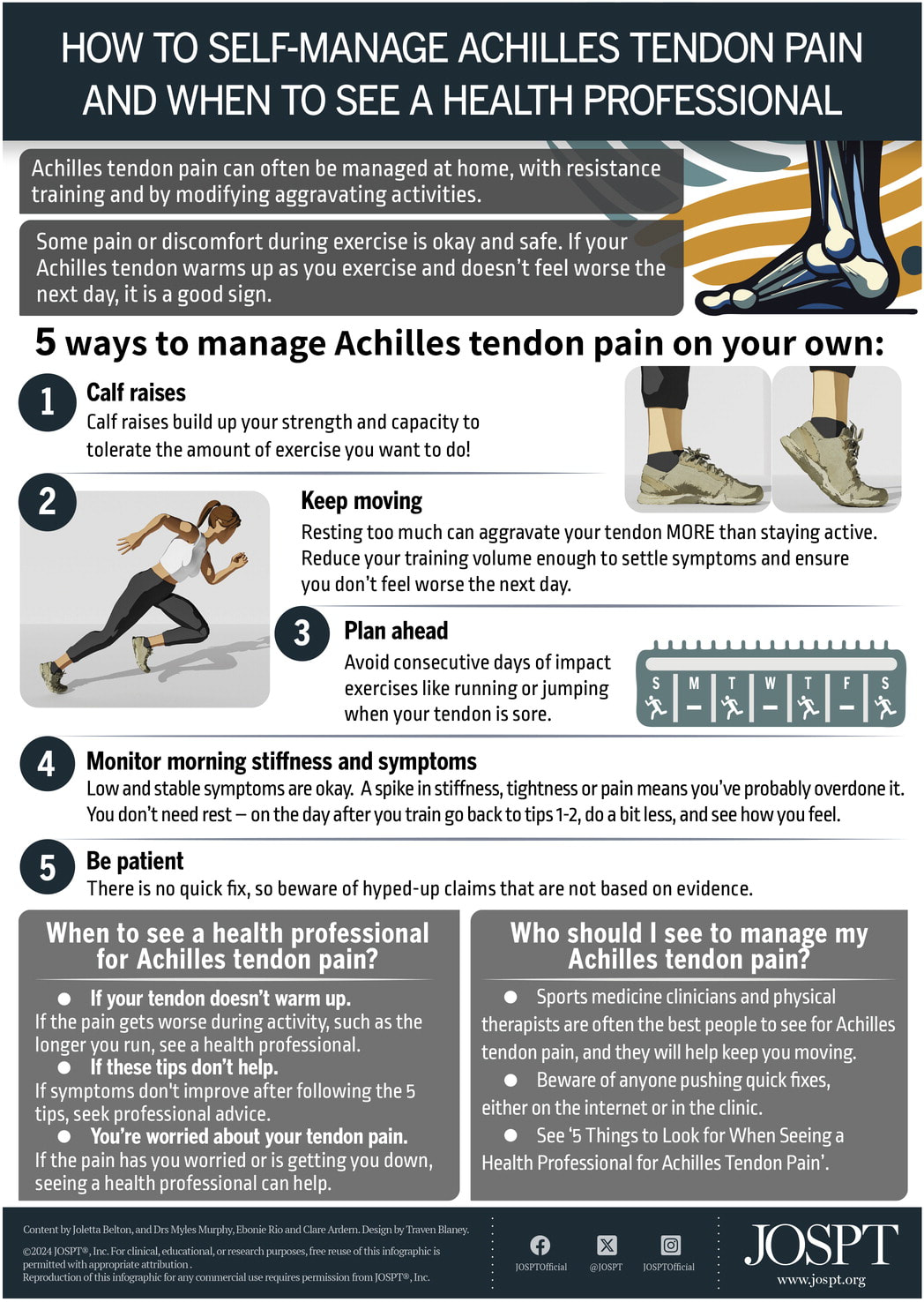
-------------------------------------------------------------------------------- 4. Animal Studies Suggest Icing Is a Double-Edged Sword While human data is scarce, the 26 animal studies in the review reveal that icing's effect is a matter of "dose and damage"—it is highly dependent on the magnitude of the injury. For large muscle injuries, the findings are predominantly negative. Icing was shown to delay muscle fiber recovery, slow regeneration, and increase the formation of scar tissue. The review illustrates this with a compelling model based on the data: for large injuries, the "regeneration" curve is visibly flattened and pushed to the right by cryotherapy, signifying a slower, less effective recovery. For minor muscle injuries, however, the story changes. In these cases, icing was found to potentially limit the expansion of the injury and accelerate regeneration. The model suggests that for minor damage, cryotherapy might contain the problem, allowing the inflammation and healing curves to resolve more quickly. This creates a dilemma: for minor muscle damage, a bit of cold may help, but for the significant tears common in sports, the same treatment appears to sabotage the processes required for a full recovery. -------------------------------------------------------------------------------- 5. The Only Undisputed Benefit Is Short-Term Pain Relief While the evidence that cryotherapy promotes healing is weak, the scientific review confirms one clear benefit: it has an undisputed analgesic (pain-relieving) effect. Applying cold numbs the affected area, which is why it feels good immediately after an injury. However, it is crucial to distinguish between numbing pain and promoting tissue regeneration. There is currently no evidence that this short-term pain relief leads to a faster or better long-term recovery. This presents the central trade-off for athletes and clinicians: using ice for immediate pain management might come at the cost of interfering with the body's biological repair processes. Based on this, the authors of the review offer a cautious recommendation. They suggest that cryotherapy may be used in the first 6 hours following an injury to reduce pain (and possibly haematoma), but advise that it "should be used with caution beyond 12 hours post-injury," as animal studies suggest it may interfere with healing. -------------------------------------------------------------------------------- Conclusion: Time to Put Old Advice on Ice? The long-standing practice of icing injuries appears to be based more on tradition than on solid human evidence. Emerging research suggests that what we've been doing for decades might not be the universally helpful intervention we once thought it was. While cryotherapy clearly has a role in managing acute pain, its continued use for accelerating healing is now highly questionable. The science is clear: our body's healing process is a controlled fire, not a wildfire to be extinguished. The question is no longer if we should use ice, but why we still do. Is it finally time to put this old advice on ice for good? -------------------------------------------------------------------------------- REF: Cryotherapy for treating soft tissue injuries in sport medicine: a critical review That sharp, nagging pain in the back of your heel—it’s often the first thing that greets you in the morning. For many, this "morning stiffness" is a frustrating daily reminder of an injury that can turn a simple walk or a beloved run into a painful ordeal. Achilles tendinopathy is common, but it's also widely misunderstood. Old advice and persistent myths can often send people down the wrong path, leading to prolonged recovery and frustration. This article cuts through the noise. Drawing on the latest physiotherapy research, we’ll reveal five surprising and counter-intuitive truths about Achilles pain. These evidence-based takeaways challenge outdated beliefs and offer a smarter, more effective path to getting back on your feet. 1. You Don't Have to Be an Elite Runner to Get It There’s a common misconception that Achilles tendinopathy is an injury reserved for serious, high-mileage athletes. While the condition is certainly prevalent in running circles, the data tells a much broader and more nuanced story. Synthesizing recent studies creates a fascinating picture: while the lifetime prevalence of the injury is a staggering 52% for long-distance runners, it’s only 6% for the general population. This seems to confirm the stereotype. However, in one study of patients in general practice, a full two-thirds of those with Achilles tendinopathy were not classified as athletes at all. Furthermore, the peak age for incidence is between 40 and 59—an age range not typically associated with peak athletic performance. The conclusion is clear: while it’s a classic “runner’s injury,” the vast majority of people who get it aren’t runners. This shift in perspective is crucial. Researchers identify the cause not as elite-level training, but as "a sudden change in stretch-shorten cycle activities"—things like walking, running, or jumping. The injury occurs when a new load exceeds the tendon’s capacity, a problem compounded by the slow adaptation of tendon compared with muscle. Understanding this helps everyone, from casual walkers to weekend warriors, recognize that a rapid increase in walking distance or starting a new fitness class can pose a risk. 2. An 'Abnormal' Scan Doesn't Mean You're Broken If you've had an ultrasound or MRI on your Achilles, the results can be alarming. Words like "thickening" or "abnormalities" sound serious and can create fear that your tendon is permanently damaged. However, a comprehensive review of imaging studies reveals one of the most counter-intuitive findings in modern sports medicine: these changes are incredibly common in people who have absolutely no pain. Research shows that up to 45% of asymptomatic people have intratendinous abnormalities, and up to 51% have tendon thickening visible on a scan. The implication here is critical: a diagnosis based only on an image can be highly misleading and may lead to unnecessary fear or interventions. A clinical diagnosis of Achilles tendinopathy is based on your history and how your tendon responds to specific loading tests (like a calf raise or a hop), not just what a scan shows. Your scan results are just one piece of a much larger puzzle, and they don't define your potential for recovery. 3. Complete Rest Is Out, and Some Pain Is Okay When something hurts, the first instinct for many is to stop moving and rest it completely. While reducing provocative activities is important, modern guidelines strongly recommend against forced, total rest for Achilles tendinopathy. In fact, it may do more harm than good. One study found that forcing athletes to completely stop their sport resulted in worse physical function at the 12-month mark compared to those who were allowed to continue their activity within acceptable pain limits. This leads to a paradigm-shifting concept in recovery: some pain during activity and exercise is acceptable. Many people experience the "warm-up phenomenon," where pain and stiffness actually improve after a few minutes of moving. During rehabilitation, a pain level of up to 5 out of 10 is often suggested as an acceptable limit for exercises. This reframes pain not as a signal of ongoing damage to be avoided at all costs, but as a guide for safely managing your activity levels. It empowers you to move away from a fear-avoidance mindset and become an active participant in your own recovery. 4. Your Mindset Matters as Much as Your Tendon For decades, tendinopathy was seen through a simple "biomedical" lens focused only on the physical tissue. Researchers now advocate for a more holistic "biopsychosocial" model, recognizing that factors beyond the tendon—our thoughts, beliefs, and emotions—play a significant role in the experience of pain and the path to recovery. Achilles tendinopathy can have a profound impact on quality of life, often rooted in a loss of identity when people are unable to participate in social and sporting activities that are important to them. The frustration and sense of limitation are a very real part of the condition, as one patient's experience illustrates: “It’s disappointing and it’s pretty frustrating, because it feels like it’s something that will never go away.” Acknowledging these psychological factors is a critical part of a successful recovery plan. Barriers to recovery often include specific, evidence-backed psychological states like kinesiophobia (a clinical fear of movement), poor outcome expectations (the belief that you may not overcome the problem), or low pain self-efficacy (feeling you are not in control of your symptoms). A modern, patient-centered approach addresses the whole person—their fears, goals, and lifestyle—not just the tissue in their heel. Conclusion The science of treating Achilles tendinopathy has evolved. What we once thought of as a simple tissue injury requiring rest is now understood as a complex condition where recovery is an active, not passive, process. The goal of modern physiotherapy is to empower you to understand your condition and its unique contributors, and to develop the confidence to self-manage your recovery through intelligent load modification and exercise. True recovery isn't just about waiting for tissue to heal; it's about challenging your beliefs about pain, engaging with your rehabilitation, and actively managing your own path back to the activities you love. Now that you know recovery is about more than just healing tissue, what is the single biggest belief about your pain that you might need to challenge first? REF: Physiotherapy management of Achilles tendinopathy
Shoulder pain is one of the most common and frustrating ailments an active person can face. Whether it’s a dull ache from rotator cuff tendinopathy (an injury to the tendons surrounding the shoulder joint, often from overuse) or a sharp pain that limits your daily activities, it can disrupt work, sleep, and exercise. For years, the path to recovery often involved a predictable sequence of scans, injections, and sometimes surgery.
But what if much of that common wisdom is wrong? A major new international clinical practice guideline, published in the Journal of Orthopaedic & Sports Physical Therapy, challenges many long-held beliefs and treatments. Based on a comprehensive review of the latest scientific evidence, it offers a new roadmap for managing shoulder pain. Here are the five most surprising and impactful takeaways that could change how you think about and manage your shoulder health. 1. You Probably Don't Need That Scan (At Least, Not Yet) When your shoulder hurts, it’s natural to want to see what’s going on inside. However, the new guidelines strongly advise against ordering diagnostic imaging tests like an MRI or ultrasound in the initial management of suspected rotator cuff tendinopathy. This new advice helps you and your doctor avoid a common, costly trap: chasing findings on a scan that may have nothing to do with your pain, which can lead to a cascade of unneeded interventions and "overmedicalisation." Instead of rushing to see inside the joint, the guidelines suggest focusing first on what has the strongest evidence for success: targeted rehabilitation. The experts state that imaging should only be considered if your symptoms fail to improve after a maximum of 12 weeks of appropriate nonsurgical management. Even more surprising is what happens when imaging is finally needed. The guideline prioritises diagnostic ultrasound over MRI. Why? Because it offers similar diagnostic accuracy for rotator cuff disorders at a lower cost. 2. Think Twice Before Getting an Injection Injections are a go-to treatment for rapid shoulder pain relief, but the new evidence suggests a more cautious approach. The guidelines now recommend against using corticosteroid (cortisone) injections as a first-line treatment. While a shot might reduce pain and disability in the short term (effects are seen for up to 8 weeks), the evidence shows that an active approach, namely a dedicated exercise program, delivers more lasting benefits. What about newer, often expensive treatments? The guideline also recommends against using Platelet-Rich Plasma (PRP) injections and Hyaluronic Acid injections as first-line treatments, citing conflicting or insufficient evidence that they work. 3. That Common Shoulder Surgery? It Might Be No Better Than a Placebo. This is perhaps the most powerful finding referenced in the new guideline. While the document focuses on non-surgical care, it highlights a critical conclusion from a previous evidence review regarding subacromial decompression surgery—one of the most common orthopaedic procedures for shoulder pain. The evidence is stark: for rotator cuff tendinopathy, this surgery provides no clinically important benefits for pain or disability when compared to a placebo (fake) surgery. This finding powerfully reinforces the guideline's theme of avoiding overmedicalisation and fundamentally questions the value of a procedure performed on countless patients who have not found relief from other treatments. 4. Exercise Is Your Best Bet—And You Can Do It at Home. Amidst the recommendations against common treatments, one approach stands out with the strongest level of supporting evidence: exercise. The guidelines state that an active rehabilitation exercise program is the recommended initial treatment for reducing pain and disability, giving it a Grade A level of evidence (the highest level of recommendation, backed by strong, consistent scientific findings). But the most empowering finding is how you can do it. According to the evidence reviewed, supervised, in-clinic exercise programs are not more effective than dedicated home-based exercise programs. This is a revolutionary finding for patients. It means you don't necessarily need to spend time and money on frequent clinic visits. The power to heal is truly in your hands, through consistent, dedicated effort at home. 5. Your Physiotherapist Should Skip These Common Treatments. Knowing what works is as important as knowing what doesn't. The new guidelines provide patients with clear, evidence-based knowledge to help them become more informed partners in their care. For the most common type of rotator cuff issue (noncalcific tendinopathy), the guideline panel explicitly recommends against using two common passive treatments during physical therapy:
If your treatment plan relies heavily on these modalities, this new evidence gives you a basis for asking your clinician about shifting the focus toward more active, exercise-based rehabilitation. Conclusion: A New Path for Shoulder Health The latest evidence on managing shoulder pain marks a clear shift in thinking. It moves away from a reliance on passive, quick-fix approaches like scans, injections, and surgery. Instead, it champions an active, patient-driven model centered on education and, most importantly, exercise. This new path emphasises that consistent, dedicated effort is the most proven way to overcome pain and restore function. Armed with this new evidence, how can you become a better advocate for your own care and partner with your clinician to build the most effective recovery plan? REFERENCE: Rotator Cuff Tendinopathy Diagnosis, Nonsurgical Medical Care, and Rehabilitation: A Clinical Practice Guideline Introduction: The Frustrating Mystery of a "Frozen" Joint
Frozen shoulder, or adhesive capsulitis, is a notoriously debilitating condition. It begins with a deep, persistent ache that soon gives way to progressive stiffness, restricting motion until even simple tasks like reaching for a shelf or putting on a coat become excruciating. For decades, it has been treated as a mysterious, localized problem—an issue confined to the capsule of the shoulder joint itself. But what if the pain and stiffness in your shoulder are not the problem, but a symptom of something much deeper? Groundbreaking research is reframing frozen shoulder entirely, suggesting it is often the "clinical expression of hormonal, metabolic, and environmental imbalances." This article explores five of the most surprising takeaways from this new, systemic perspective on what's really happening when a shoulder "freezes." Your Shoulder Is the Symptom, Not the Source It’s a systemic issue, not just a local one. The first major paradigm shift in understanding frozen shoulder is moving away from the idea that it’s purely a mechanical joint problem. The traditional view of a simple inflammation and scarring of the joint capsule is now considered incomplete. Instead, research paints a picture of frozen shoulder as a systemic disorder involving widespread, low-grade inflammation and dysfunction across the body's endocrine (hormonal), immune, and metabolic systems. The shoulder joint, with its unique anatomy and blood supply, becomes the target where these system-wide issues finally manifest as pain, fibrosis, and stiffness. It is the end point of a cascade of problems, not the starting point. This perspective helps explain why it's so strongly linked to other systemic conditions and why purely local treatments often fall short. "FS is not merely a localized joint pathology but a systemic disorder requiring integrative clinical strategies..." The Estrogen Story Is More Complicated Than Menopause It’s not just estrogen deficiency, but estrogen resistance. The strong link between frozen shoulder and women in their 40s and 50s has long pointed to the hormonal shifts of perimenopause and menopause. While declining estrogen levels are certainly a major factor, the story is more nuanced than simple deficiency. The new research highlights the crucial concepts of "estrogen resistance" and "receptor-level interference." This is a state where estrogen may still be present in the bloodstream, but the body's tissues—including the cells in the shoulder capsule—have lost their ability to hear its signals and respond properly. This resistance can be driven by a number of factors that interfere with the estrogen receptors themselves.
This is a critical insight because it helps explain why frozen shoulder can affect women who are not yet postmenopausal and underscores that the hormonal environment is shaped by much more than just age. Your Modern Lifestyle Might Be the Root Cause The "Lifestyle Hypothesis" connects daily habits to chronic inflammation. If systemic inflammation and metabolic dysfunction are setting the stage for frozen shoulder, what’s causing them? The "Lifestyle Hypothesis" proposes that the underlying driver is often modern life itself. Common daily behaviors can create and sustain a state of chronic low-grade inflammation (LGI), which acts as the foundation for developing the condition. These lifestyle factors are not dramatic, one-time events, but rather the cumulative effect of daily habits:
In essence, this hypothesis suggests that the daily, seemingly minor choices—the processed snack, the extra hour of screen time, the stressful commute—accumulate to create the exact inflammatory and metabolic storm in which a condition like frozen shoulder can thrive. This constant, low-level activation creates a vicious cycle: chronic inflammation causes insulin and estrogen resistance, and these resistance states, in turn, fuel more inflammation, locking the body in a self-perpetuating loop. It Has Hidden Links to Your Thyroid and Blood Sugar FS is strongly associated with other metabolic conditions. The systemic nature of frozen shoulder is starkly illustrated by its strong connection to other health conditions, some of which are often overlooked in a standard orthopedic evaluation. Beyond the well-known link to diabetes, research confirms significant associations with thyroid and metabolic health. A recent systematic review and meta-analysis confirmed that both overt and subclinical hypothyroidism significantly increase the risk of developing frozen shoulder. Furthermore, a type of genetic study known as Mendelian randomization provides strong support that hypothyroidism can be a direct cause. Crucially, researchers have found that hypothyroidism can create a state of functional estrogen resistance by altering key proteins that transport sex hormones and by inducing insulin resistance—brilliantly connecting this finding back to the central theme of estrogen signaling failure. Similarly, the metabolic connection is undeniable. Meta-analyses consistently find that patients with frozen shoulder have elevated levels of HbA1c—a blood test that shows your average blood sugar levels over the past two to three months—and high cholesterol. This takeaway is critical because it suggests that a complete evaluation for someone with frozen shoulder should include screening for these conditions. Managing underlying thyroid or blood sugar issues may be a crucial, and often missing, component of a successful recovery. Healing May Require a Whole-Body Approach The future of treatment goes beyond injections and physiotherapy. If frozen shoulder is a systemic problem, then the solution must also be systemic. This new understanding implies that effective, long-term treatment needs to go beyond conventional approaches like corticosteroid injections and physiotherapy to address the root causes of inflammation and metabolic imbalance. This new model shifts the focus from passively receiving treatment to actively rebuilding the body's systemic health, turning a shoulder problem into an opportunity for whole-body wellness. Key components of this integrative strategy include:
As researchers put it, the path to healing is about restoring balance on a much broader scale. "...the future of FS management lies not only in better understanding the joint capsule—but in restoring systemic balance at the intersection of hormones, metabolism, immunity, and environment." Conclusion: Unlocking the Shoulder by Rebalancing the BodyThe emerging science of frozen shoulder is transforming our understanding of this painful and limiting condition. It is moving from being seen as an isolated orthopedic issue to being recognized as a clear signal of deeper, systemic imbalance across our hormonal, metabolic, and immune systems. This new perspective offers not only better treatment strategies but also a profound opportunity for improving overall health. This paradigm shift leaves us with a compelling question: Could listening to the message from your shoulder be the first step toward improving your overall metabolic, hormonal, and systemic health for years to come? REF: Frozen Shoulder as a Systemic Immunometabolic Disorder: The Roles of Estrogen, Thyroid Dysfunction, Endothelial Health, Lifestyle, and Clinical Implications
|
Archives
April 2026
Categories
All
|
RSS Feed